Membrane morphogenesis
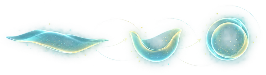
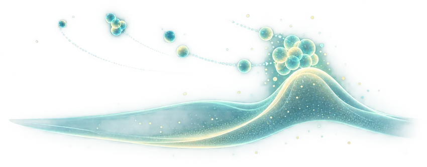
How does a flat membrane curve into a new cellular compartment? We combine microscope images with physical models to explain changes in membrane shape.
\n
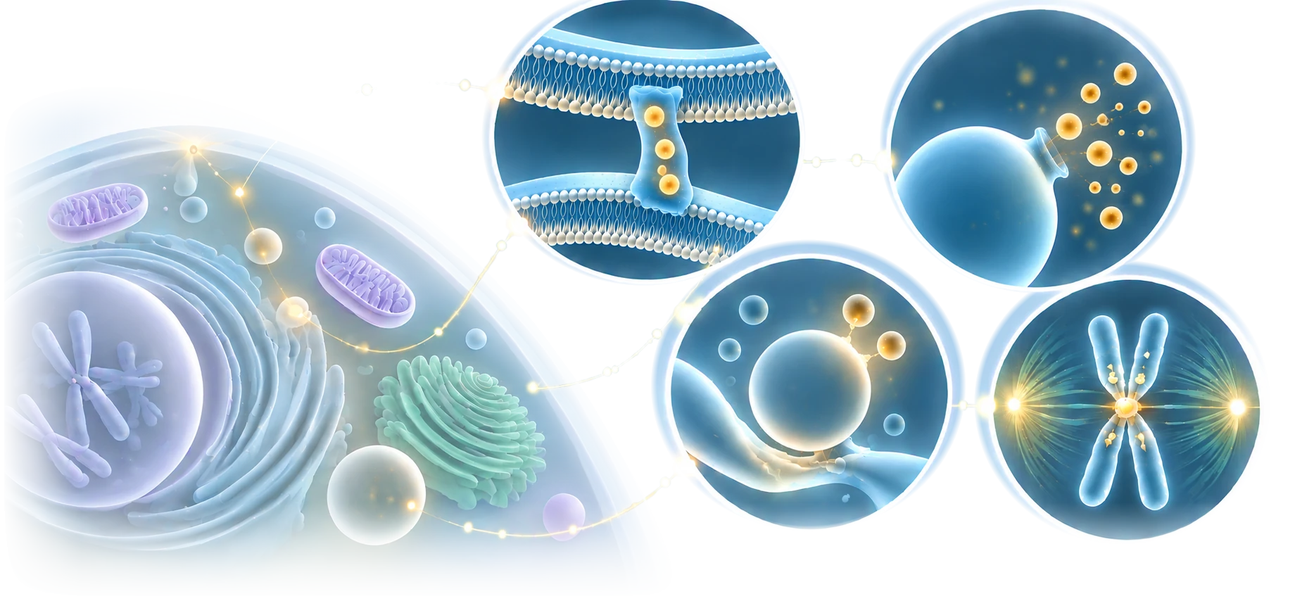
Cells are always changing. How do they keep working reliably? I study how membranes are built, moved and recycled, and how chromosomes are passed accurately to new cells.

From molecular interactions to the architecture of cells, physical principles connect biological function across scales.
Two seemingly different biological questions, connected by the physics of dynamic organization.
How cells keep their inner structures working while membranes are built, moved and recycled.
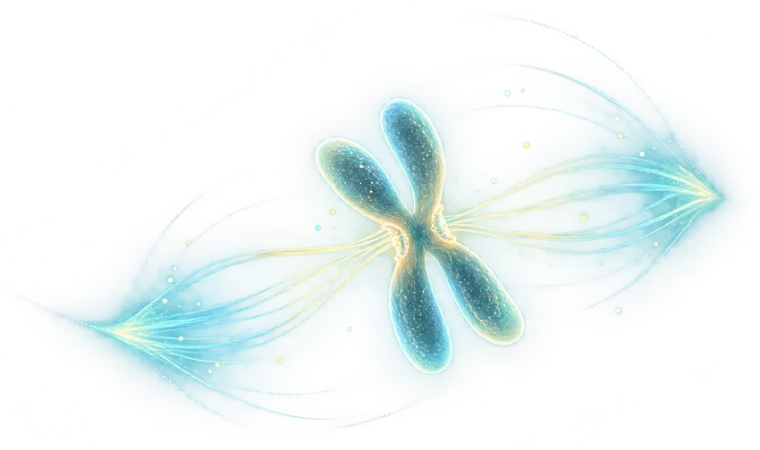
How cells organize and separate chromosomes so each new cell receives the right genetic information.
How can biological structures remain reliable yet adapt, reorganize, and respond to change? Homeostasis and fidelity share this underlying challenge.
Maintain function despite fluctuations
Reconfigure structure when needed
Create architecture from local interactions
Act at the right place and time
We combine simple physical models, computer simulations of moving molecules and experiments to study the same problem from different angles.
How does a flat membrane curve into a new cellular compartment? We combine microscope images with physical models to explain changes in membrane shape.
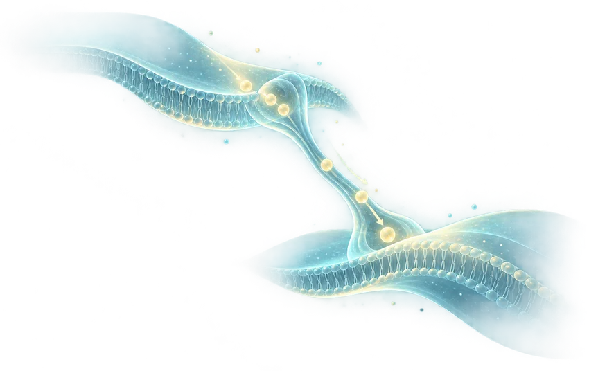
How do cellular compartments share the lipids needed to build their membranes? We use molecular simulations to study proteins that bridge nearby membranes.
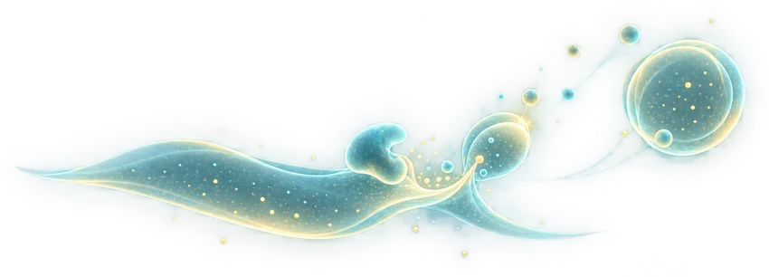
How does a cell break down the right membrane at the right time? We study how membranes are recognized, become ready to fuse and are safely recycled.
What happens when many proteins gather in one place? We study how droplet-like assemblies help cells recycle materials, bring membranes together and change shape.
Why does a chromosome need the right shape to separate correctly? Physical models show how long DNA molecules can be packed and pulled apart during cell division.
How does a dividing cell pull chromosomes apart without mistakes? We study the connections that hold chromosomes, the signals that correct errors and the fibers that move them.
My work began with theoretical physics and mathematical models of chromosome organization and membrane remodeling. I now combine molecular simulations with experimental studies to investigate the mechanisms behind these processes.
Physical models help us explore how membranes bend, long DNA molecules fold, and proteins come together to build functional structures.
Molecular dynamics is a computer method for following how atoms or groups of atoms move. We use it to study changes in molecular shape, how molecules assemble and interact, and how forces at the molecular scale give rise to biological functions.
Imaging, structural biology and biochemical measurements help test predictions and connect molecular mechanisms to cellular function.
Eight research highlights spanning mathematical models, molecular mechanisms and cellular organization. Find additional papers in the individual research pages.
Structural and functional analyses explain how a lipase recognizes a membrane and becomes active during breakdown.
Membrane curvature · lipid degradationThe preprint investigates how phosphorylation regulates the movement of lipids through a membrane-bridging protein.
Phosphorylation · lipid transferThe preprint examines a shell-like higher-order arrangement of proteins that build the kinetochore.
Kinetochore architecture · multiscale simulationPhosphorylation changes the properties of p62 condensates and affects how unwanted proteins are removed.
Condensates · selective autophagyA protein assembly can generate local osmotic effects that drive outward protrusions of the cell membrane.
Membrane blebbing · physical mechanismsThree-dimensional measurements and physical modeling reveal characteristic shapes formed as autophagosomes grow.
Membrane geometry · mathematical theoryMeasurements of individual nucleosomes clarify how condensin activity and contacts within chromatin shape mitotic chromosomes.
Chromatin dynamics · mesoscale simulationA polymer model connects the shape of intertwined chromosomes with how quickly they separate.
Polymer physics · chromosome separationMoving from physical models of organization toward experimentally testable molecular mechanisms.
Polymer simulations establish physical models linking chromosome geometry to segregation speed.
Mathematical modeling connects chromosome shaping with segregation.
Membrane mechanics explains autophagosome shape transitions.
Quantitative microscopy and simulations connect scales.
Membrane turnover, lipid flow and kinetochore architecture.
I am a computational and theoretical biophysicist interested in how living matter self-organizes. Trained in theoretical physics, I use mathematical modeling and molecular dynamics to understand how cells build membranes and establish the physical structures needed for accurate chromosome inheritance.
For scientific discussions, collaborations and inquiries.