Explaining observed 3D shapes with physical models
Can measurements of actual membranes test a model of how they bend?
How it works
A single microscope picture cannot show the full shape of a membrane: the same structure can look very different from different angles. We therefore reconstruct entire membranes in three dimensions and compare their measured shapes with predictions from physical models.
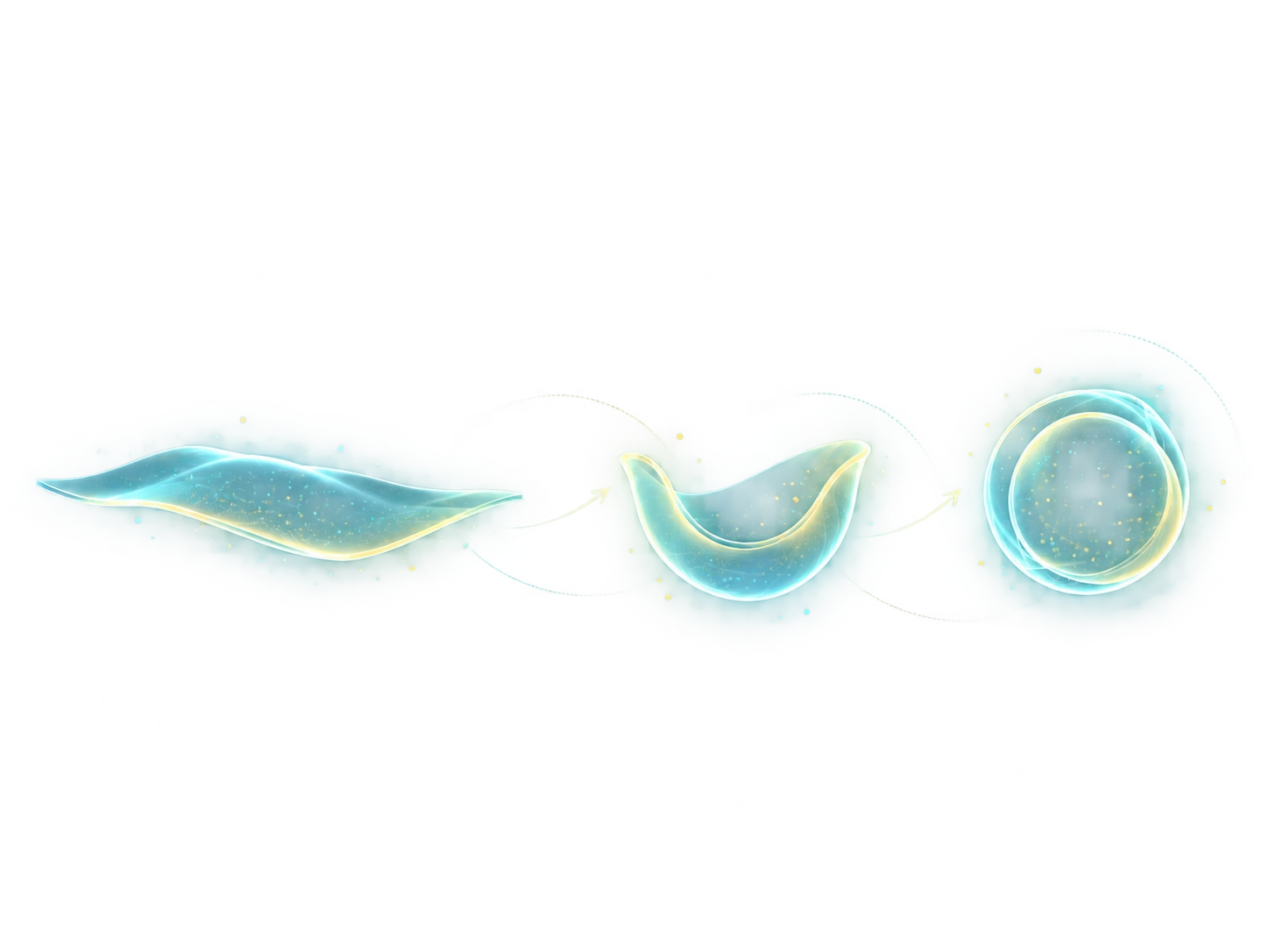
In a 2024 study, we compared 117 three-dimensional structures of forming autophagosomes. Many were not simple round bowls. Instead, they resembled elongated cups whose open edges curved outward.
We then calculated the energy needed to bend membranes into different shapes. Comparing these predictions with real structures helps explain why particular forms appear, rather than simply describing how they look.
What we want to learn next
Published researchWhere the observed shapes differ from predictions, we can ask whether membrane-bending proteins or differences in lipid distribution between the two sides of the membrane are responsible.
Related publications & press releases
Three-dimensional measurements and physical modeling reveal characteristic shapes formed as autophagosomes grow.